
Tibial Plateau Fracture: From Surgery to Recovery
Case study: a complex lateral tibial plateau fracture treated by Dr. Andrei Ursache using a single-implant technique (LISS plate), followed by complete recovery within one year.
Surgery, and recovery afterward, for a lateral tibial plateau fracture can be considerably more difficult when the injury is complex. With the help of new surgical techniques and technologically advanced implant systems, it is now possible to perform an operation that fixes the fractured bone correctly and quickly, giving the patient a real advantage when it comes to recovery.
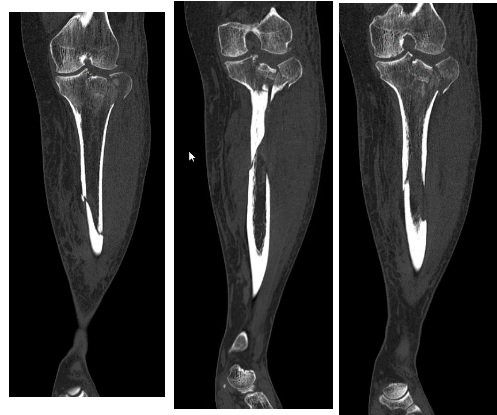
A 50-year-old patient came to Clinica Provita following a motorcycle accident, with a depressed lateral tibial plateau fracture extending into the shaft of the tibia. The patient was taken into care by Dr. Andrei Ursache, senior consultant in Orthopedics and Traumatology.
His medical history included an arthroscopic surgery five years earlier, involving chondroplasty for cartilage defects on the medial femoral condyle and the femoropatellar joint.
Surgery for the lateral tibial plateau fracture
Dr. Andrei Ursache took into account the specific features of the fracture: the postero-lateral position of the depressed tibial plateau segment, and the oblique-spiral extension of the fracture into the shaft, reaching down to the middle third of the bone.
The surgical solution therefore had to achieve:
Open reduction and internal fixation of the tibial plateau fracture;
Reduction (open or closed) of the diaphyseal fracture;
Fixation of the diaphyseal fracture;
Internal fixation that would allow joint mobilization immediately after surgery;
As short a period as possible of restricted weight-bearing on the lower limb.
The implant – a 13-screw LISS plate
The surgeon chose to use a single implant capable of providing absolute stability for the tibial plateau fracture. With several treatment options available, he opted for a single-implant surgical technique. "The solution we chose was to use a single implant, one that would make it possible to achieve absolute stability for the tibial plateau fracture and relative stability for the diaphyseal fracture," says Dr. Ursache.
This approach allowed the patient to be positioned on the operating table only once, lying on his back, with two relatively limited surgical approaches totaling less than 10 cm.
Dr. Ursache explained the requirements the implant needed to meet: "The fixation plate had to support the postero-lateral fragment of the tibial plateau, provide angular stability for the proximal screws, be long enough to extend at least 3 screws beyond the distal fracture site, and offer a working length suited to elastic fixation along the diaphyseal fracture line."
Following the preoperative plan, the surgeon chose the 13-screw LISS plate as the implant, since it met all the required characteristics, while also being made of titanium — a material with excellent biological tolerance — and featuring an instrumentation set that is very surgeon-friendly during implantation.
Dr. Ursache notes that another challenge was approaching the postero-lateral fracture through an antero-lateral approach, contrary to the usual principle of operating "where the fracture is." Analysis of the CT images made it possible to identify an antero-lateral fracture line, which Dr. Andrei Ursache used intraoperatively to reach the depressed fragment.
Post-operative recovery
The goal of modern internal fracture fixation is immediate joint mobilization and, whenever possible, the fastest possible return to weight-bearing on the limb. Modern traumatology follows a guiding principle: "Movement is life, and life is movement." The surgery proceeded under favorable conditions, with all planned objectives achieved. Joint mobilization began immediately after the operation. Partial weight-bearing on the lower limb started at 8 weeks, and full weight-bearing at 16 weeks.
Six months after surgery, the patient reported clicking sensations when moving the left knee, unaccompanied by pain, with a palpable correlate at the level of the kneecap.
To investigate the source of these clicking sensations, and to check for any irregularities resulting from imperfect healing of the lateral tibial plateau, arthroscopy of the left knee was decided upon and performed. Inspection showed that the lateral tibial plateau had healed perfectly, but revealed a recurrence of osteochondral lesions on the medial femoral condyle and the femoral trochlea. Microfracture chondroplasty was performed on the lesions, followed three weeks later by intra-articular treatment with PRP + Hyaluronate (Cellular Matrix, Regenlab).
One year after surgery, the patient walks without pain and is even able to play sports, albeit at a moderate level of impact.