Ronda Medical
Finceramica

CustomBone Service™ — Patient-specific cranioplasty implant

The only patient-specific implants made of bioceramic coated with hydroxyapatite — starting from the patient’s CT scan, they reconstruct large and complex cranial defects with a bio-mimetic material that behaves like natural bone and promotes bone regeneration.

Product gallery
CustomBone Service™ — porous hydroxyapatite cranioplasty implant with a bio-mimetic surface (Finceramica material)
CustomBone Service™ — 3D model of the patient’s skull with the custom implant positioned over the defect (Finceramica material)
CustomBone Service™ — detail of the interconnected porosity of the hydroxyapatite, which promotes osseointegration (Finceramica material)
Porous hydroxyapatiteMaterial
~70%HA in human bone
~2 monthsDelivery
2 (+ spare)Implants supplied
Clinical description

CustomBone Service™ represents the only patient-specific implants made of bioceramic coated with hydroxyapatite, developed by Finceramica for the reconstruction of large and complex cranial defects. Unlike conventional techniques, the implant targets not only the aesthetic outcome but also natural bone regeneration, with neurological and psychological benefits for the patient.

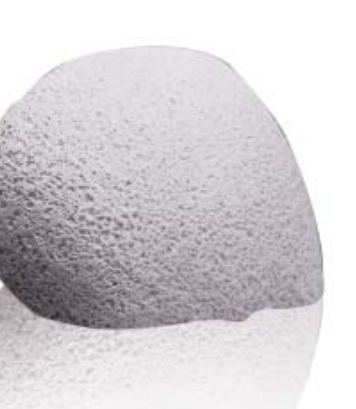
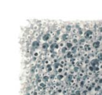
The material is a bio-mimetic ceramic based on macro- and microporous hydroxyapatite — the same component that makes up about 70% of human bone. The interconnected porosity promotes rapid cellular colonization, deposition of new bone, and neovascularization of the implant, supporting osseointegration through direct contact with the adjacent vital bone. The material is fully biocompatible, with no risk of viral transmission, and in the event of subsequent trauma it behaves like natural bone. Owing to the manufacturing process, the hydroxyapatite crystals are more resistant to osteoclastic activity (less osteolysis than an autologous graft), undergoing slow, physiological remodeling.

The CustomBone Service™ process starts from the raw data of the patient’s computed tomography (performed according to a dedicated protocol); through 3D reconstruction and stereolithography, a synthetic model of the skull is obtained, then a prototype of the implant, validated together with the surgeon before finalization. The final implant is prepared, refined, inspected, and sterilized, and is delivered in approximately 2 months. Two implants are supplied (primary + spare), to allow an immediate replacement in the event of a second trauma. Fixation is usually performed with simple sutures — rigid fixation methods, which could fracture the porous hydroxyapatite, are not used.

Indications
  • Reconstruction of large and complex cranial defects after decompressive craniectomy.
  • Cranial fractures caused by trauma.
  • Cranial defects after tumor resection.
  • Resorption of an autologous bone graft.
  • Infection or rejection of a previously applied material.
Key features

Bio-mimetic porous hydroxyapatite

The same chemical composition as the mineral component of human bone (~70% of bone).

Custom-made from CT

Implant produced individually from the patient’s CT data: CT → 3D model → validated prototype → final implant.

True osseointegration

Interconnected porosity that promotes cellular colonization, bone deposition, and neovascularization.

Behaves like natural bone

In the event of subsequent trauma it can show biological activity and self-healing, like native bone.

Resistant to osteolysis

HA crystals more resistant to osteoclastic activity than an autologous graft, with slow physiological remodeling.

Simplified procedure

Fixation with simple sutures, natural aesthetic result; a second (spare) implant is included for a possible second trauma.

Technical specifications
Type
Patient-specific (custom-made) cranioplasty implant, made of porous hydroxyapatite
Material
Bio-mimetic ceramic based on macro- and microporous hydroxyapatite
Production
From the patient’s CT scan (dedicated protocol) → stereolithographic 3D model → prototype validated with the surgeon → final implant
Fixation
Simple sutures; direct contact with vital bone (no rigid fixation)
Delivery
Approximately 2 months; a primary implant and a spare are supplied
Minimum age
Not recommended for large/complex defects in children under 7 years (case-by-case for small defects)
Osseointegration
Begins after contact with vital bone; visible on CT at 6–8 months postoperatively
Clinical evidence

A selection of publications and studies that mention use of the system. Summaries are indicative; for details, consult the original source.

Long-Term Follow-Up of Custom-Made Porous Hydroxyapatite Cranioplasties: Analysis of Infections in Adult and Pediatric Patients

Mannella F.C., Faedo F., Fumagalli M., Norata G.D., Zaed I., Servadei F. · Journal of Clinical Medicine, 2024; 13:1133

Post-marketing surveillance study in 687 patients (including 80 pediatric, under 14 years), from 26 neurosurgery centers in five European countries, which analyzed complications and infections after cranioplasty with custom-made porous hydroxyapatite (PHA) implants.

Infections were the main complication — 6% of implants (10% in pediatric vs. 5.4% in adult patients); explantation was required in 4.4% of patients. Bifrontal decompression was associated with increased risk (12.5% vs. 5.1%).
View source
Certifications & compliance
CE marking